Rib Cage Muscles Anatomy : Claire Lordon Design: Écorché Rib Cage to Humerus Bone and ... / They are more involved in forced expiration and coughing to forcibly shrink the chest and.
Rib Cage Muscles Anatomy : Claire Lordon Design: Écorché Rib Cage to Humerus Bone and ... / They are more involved in forced expiration and coughing to forcibly shrink the chest and.. Structure of a typical rib: Ribs & thoracic cage muscles attachments. See more ideas about anatomy, anatomy study, rib cage anatomy. Learn anatomy faster and remember everything you learn. Learn about ribs muscle with free interactive flashcards.
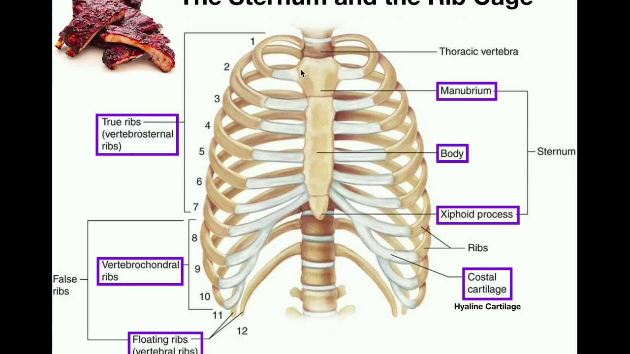
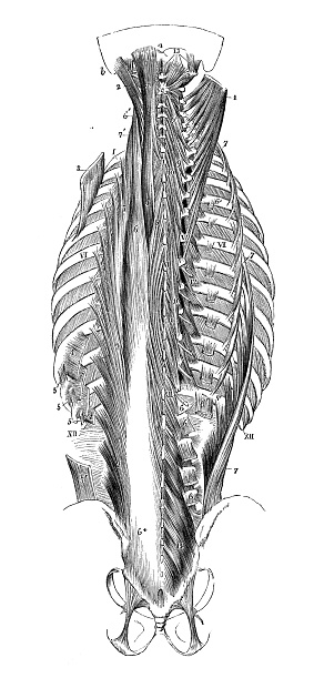
While muscle spasms may occur over the entire body, muscle spasms under the rib cage may be cause for concern as they might be an indication of serious medical conditions. The fibers attach to the rib cage and the pubis of the hip bones. Anatomy of a human body we study anatomy. 1887 human anatomy print of the rib cage and sternum. The rib cage, shaped in a mild cone shape and more flexible than most bone sets, is made up of varying elements such as the thoracic vertebra, 12 equally paired ribs, costal cartilage, and held together anteriorly by the sternum.
Anatomy | The Sternum, Rib Cage, & Vertebrae - YouTube from i.ytimg.com The fibers attach to the rib cage and the pubis of the hip bones. Seventeen muscles attach to the scapula, and it articulates with the clavicle to form the shoulder girdle or pectoral girdle, which supports movements. Tendons attach the muscles to each other. In most tetrapods, ribs surround the chest, enabling the lungs to expand and thus facilitate breathing by expanding the chest cavity. Переглядів 46 тис.9 років тому. Check out our muscle anatomy reference charts to learn faster! This is a stereogram, to be viewed in crossview technique. The rib cage, shaped in a mild cone shape and more flexible than most bone sets, is made up of varying elements such as the thoracic vertebra, 12 equally paired ribs, costal cartilage, and held together anteriorly by the sternum.
Muscles of thoracic age are the intercostals (external, internal and innermost), subcostals, and.
The muscles that affect the knee's movement run along the thigh and calf. The ribs are curved, flat bones which form the majority of the thoracic cage. Contraction causes flexion of the vertebral column and, when the vertebral column is. Structure of a typical rib: Contributing to their role in protecting the internal thoracic organs. For example, flexor, extensor, adductor and abductor are names associated with the action of the muscle. Illustration of rib cage, demonstrating ribs and connection through cartilage to sternum. Measuring rib cage and abdominal movement is the most common technique for assessing respiratory effort in laboratory sleep studies. Rib cage anatomy and breathing. Various skeletal muscles are attached to the rib cage. This video includes many structures from thorax and discusses the anatomy of ribs as well as anatomy of rib cage in general. This cage protects vital organs and is essential for creating negative pressure to inflate lungs. Ribs & thoracic cage muscles attachments.
During normal breathing, contraction of the major inspiratory muscle, the diaphragm, produces both rib cage expansion and a downward movement of the diaphragm. Everyone has nice muscles in ct scanning! Muscular system anatomy:muscles of the thoracic cage torso model description. The cartilages of three other ribs are connected with. Tendons attach the muscles to each other.
Antique Illustration Of Human Body Anatomy Neck Spine Rib ... from media.istockphoto.com Rib cage diagram with organs. In most tetrapods, ribs surround the chest, enabling the lungs to expand and thus facilitate breathing by expanding the chest cavity. Переглядів 46 тис.9 років тому. The fibers attach to the rib cage and the pubis of the hip bones. Anatomical illustration, images of the human body, pepin press. Illustration of thoracic vertebrae showing vertebral body, pedicles, facets, transverse process, rib. Each rib articulates posteriorly with the vertebral column. The muscles that affect the knee's movement run along the thigh and calf.
Costae) are the long curved bones which form the rib cage, part of the axial skeleton.
It provides a strong framework onto which the muscles of the cramps in ribcage are often observed in those who strain or overwork their upper body. Each rib articulates posteriorly with the vertebral column. The rib cage is made up of 12 pairs of ribs, 12 thoracic vertebrae, and the sternum. Top suggestions for rib cage anatomy muscles. Everyone has nice muscles in ct scanning! Muscles of thoracic age are the intercostals (external, internal and innermost), subcostals, and. Various skeletal muscles are attached to the rib cage. Thoracic vertebral column twelve pairs of ribs: Contraction causes flexion of the vertebral column and, when the vertebral column is. This cage protects vital organs and is essential for creating negative pressure to inflate lungs. In most tetrapods, ribs surround the chest, enabling the lungs to expand and thus facilitate breathing by expanding the chest cavity. In vertebrate anatomy, ribs (latin: Learn anatomy faster and remember everything you learn.
Everyone has nice muscles in ct scanning! Rib cage diagram with organs. Muscle spasms located in the rib cage are often observed in people who strain or overwork their upper body muscles. See more ideas about anatomy, anatomy study, rib cage anatomy. Some extend from above and draw the.
AnatomyTools from www.anatomytools.com All muscles that are attached to the human rib cage have the. The fibers attach to the rib cage and the pubis of the hip bones. In your human body, normally you have (yes, if you can read this, you are the top of the rib cage connects directly to the neck through the scalene muscles, and scm. They are attached to the femur (thighbone), tibia (shinbone), and fibula (calf bone) by fibrous tissues called ligaments. Ribs & thoracic cage muscles attachments. Contributing to their role in protecting the internal thoracic organs. Check out our muscle anatomy reference charts to learn faster! The ribcage is made to be flexible and springy so the lungs can fill and deflate easily.
During normal breathing, contraction of the major inspiratory muscle, the diaphragm, produces both rib cage expansion and a downward movement of the diaphragm.
The ribs are a set of twelve paired bones which form the protective 'cage' of the thorax. Thoracic vertebral column twelve pairs of ribs: The neck muscles (and neck anatomy on the whole) are responsible for head movement, stabilizing the upper region of the body, assisting in swallowing, helping to elevate the rib cage during inhalation, and more. Muscle spasms located in the rib cage are often observed in people who strain or overwork their upper body muscles. During normal breathing, contraction of the major inspiratory muscle, the diaphragm, produces both rib cage expansion and a downward movement of the diaphragm. Muscles are often named for their primary action. Collectively referred to as the rib cage costal cartilages sternum. Anatomy of a human body we study anatomy. The thorax is anatomical structure supported by a skeletal framework (thoracic cage) and the ribs on both the sides complete the cage. See more ideas about anatomy, anatomy study, rib cage anatomy. The cartilages of three other ribs are connected with. The ribcage is made to be flexible and springy so the lungs can fill and deflate easily. All muscles that are attached to the human rib cage have the.
Muscular system anatomy:muscles of the thoracic cage torso model description rib cage muscles. The neck muscles (and neck anatomy on the whole) are responsible for head movement, stabilizing the upper region of the body, assisting in swallowing, helping to elevate the rib cage during inhalation, and more.